
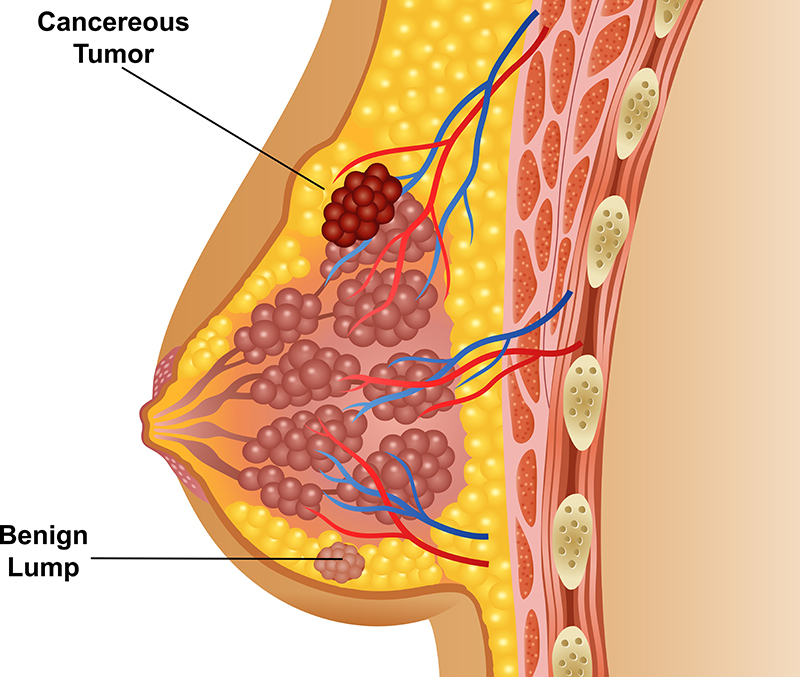
Benign Tumours of the Breast
Benign breast tumor treatment in Chennai is administered with excellent care in our clinic. As the condition is harmless, we emphasize on frequent consultation and self-examination procedures. There are possibilities of non- cancerous cells to transform into cancerous cells.
Our specialist suggests effective ways to monitor the changes in the body. It can help to commence treatments at an early stage, if necessary. His wide medical competence aids in developing the clinic as the best place to receive breast tumor surgery in Chennai.
Non-cancerous breast abnormalities are considered as benign breast conditions. They are not life-threatening but can cause pain and discomfort in certain individuals. Certain benign conditions can develop into cancerous growths.
Anatomy: The breast constitutes of glandular and stromal tissues. The milk-producing lobules and ducts are contained in the glandular tissues while the fibrous and fatty connective tissues are in the stromal tissues. Abnormal changes to any of these portions can lead to benign breast conditions.
Benign breast conditions are broadly classified as:
- Mastitis – The defect is associated with fever, warmth in the area, induration, localized erythema, tenderness and swelling.
- Breast mass – The mass is firm and varies in shape and size. They are usually located in the upper right quadrant (of the breast). Patients may experience pain with painless palpation. Changes may be noted such as asymmetry, inversion or nipple discharge, skin tethering and retraction, peau d’orange or skin inflammation, and axillary lymphadenopathy.
- Breast abscess – These usually occur in the periareolar or areolar region. Swelling is noted with no palpable mass. Patient suffers from fever, discharge from the nipple, breast appears erythematous, induration, warmth in the area, oedema and tenderness.
Causes
- Fibroadenoma – This is one of the most common causes of mass formation in women < 25 years of age. The benign malignancy arises from the terminal duct lobular unit and can even appear as multiple masses. They are firm, smooth, mobile and rubbery and are painless. Their size ranges from 1 cm to about 5 cm. They can enlarge causing changes in the skin and contours of the breast. A well-hypoechoic homogenous mass (about 1cm to 20 cm in diameter) is detected on ultrasonography.
- Fibrocystic changes – Cysts are formed due to dilated lobules of the breast. The main cause is associated to changes in menstrual cycle. The women developing fibrocystic changes are aged anywhere from 35 to 50 years. The cyst could also rupture causing inflammation and scarring. Fibrotic changes are firmness and rubbery consistency.
- Hyperplasia – Cells that line the ducts or lobules experience an overgrowth. Most women have mild hyperplasia. It is only when there is a presence of atypical hyperplasia, that it is considered as malignant.
- Phyllodes tumour – It is a giant fibroadenoma also known as cystosarcoma phyllodes. This commonly occurs in women aged between 40 years and 60 years. The presence of a large, firm, single breast nodule is noted.
- Mastitis – Occurring in lactating women, mastitis originates while weaning off breast feeding or after 6 weeks of postpartum. Staphylococcus aureus is the most common cause of the condition.
- Breast abscess – Methicillin-resistant S.aureus along with streptococcal species are responsible for puerperal breast abscesses. The condition leads to non-lactation and diabetes in women. Smoking is also a risk factor.
- Papillary adenoma of the nipple – The erosive adenomatosis of the nipple arises from the terminal lactiferous ducts. The women affected usually range from the ages of 40 years to 50 years.
- Adenosis – When the number of glands increases, it is known as adenosis.
Diagnosis and Testing
Imaging studies – Ultrasonography differentiates between the solid and cystic structures. Simple cysts are round and oval whereas complex cysts are solid lobulations containing internal debris.
Treatment
Antibiotic therapy with Nafcillin, Vanomycin and Clindamycin will reduce inflammation and subsequent complications.
Ask doctor
Testimonials
 Very practical approach to my dads gallbladder stone problem .. Surgery was explained well by diagrams and he performed the surgery by key holes which made it pain free for my dad . I had consulted many in the last 1 month including
Subramanian
Read More The doctor was helpful. He worked with me to select the best option for treatment and helped finalize treatment. He ensured that my appointment went ahead as planned and followed up rigorously post op too. Scar was a bit larger
Mehul Kain
Read More The doctor was helpful. He worked with me to select the best option for treatment and helped finalize treatment. He ensured that my appointment went ahead as planned and followed up rigorously post op too. Scar was a bit larger

